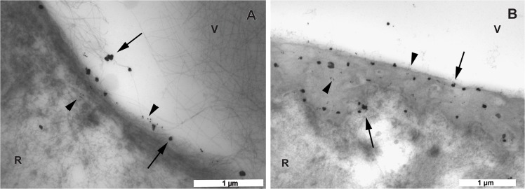
Fig 8. Transmission electron microscopic images of the vitreoretinal interface double labeled with antibodies against type IV and VI collagens.
The large labels are gold particles with silver enhancement representing type VI collagen (arrows). The small labels are 15 nm gold particles representing type IV collagen (arrow heads). A. In the eye of the 56 year-old donor, the equatorial area exhibits a linear distribution pattern of both type IV and VI collagens. Note that type VI collagen can also be seen on the vitreous fibrils. B. In the eye of the 35 year-old donor, towards the posterior pole, both type IV and VI collagens become diffusely distributed over the entire thickness of the ILM. A, B bar = 1 μm. V = vitreous; R = retina.

