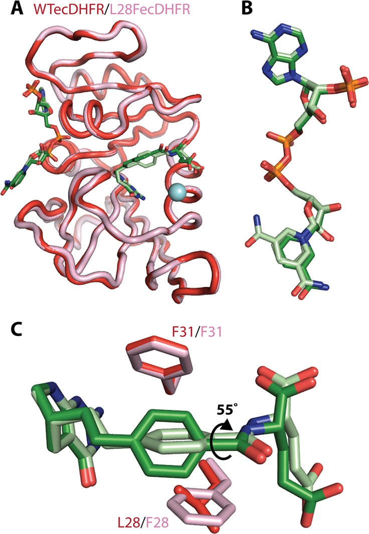
Figure 2.

(A) Overlay of the WT E:ddTHF:NADP+ (red) and L28F E:ddTHF:NADP+ (pink) crystal structures. The backbone is shown as a cartoon and the ddTHF and NADP+ ligands as sticks. The site of the L28F mutation is shown as a blue sphere. (B) Conformation of NADP+ in WT E:ddTHF:NADP+ (dark green) and L28F E:ddTHF:NADP+(pale green) complexes. In both structures, the nicotinamide ring projects into the solvent and packs against a neighboring molecule in the crystal lattice. The carboxamide moiety is found in two orientations (related by 180° flips about the axis of the nicotinamide ring) in the L28F E:ddTHF:NADP+ structure. (C) The benzene ring of the of the para-ethyl benzoyl glutamic acid tail (PEBA) of ddTHF is rotated 55° around its C1′-C4′ axis in L28F E:ddTHF:NADP+ (pale green, pink) relative to its orientation in the WT complex (dark colors), altering its contacts with F31 and aliphatic side chains lining the binding pocket.
