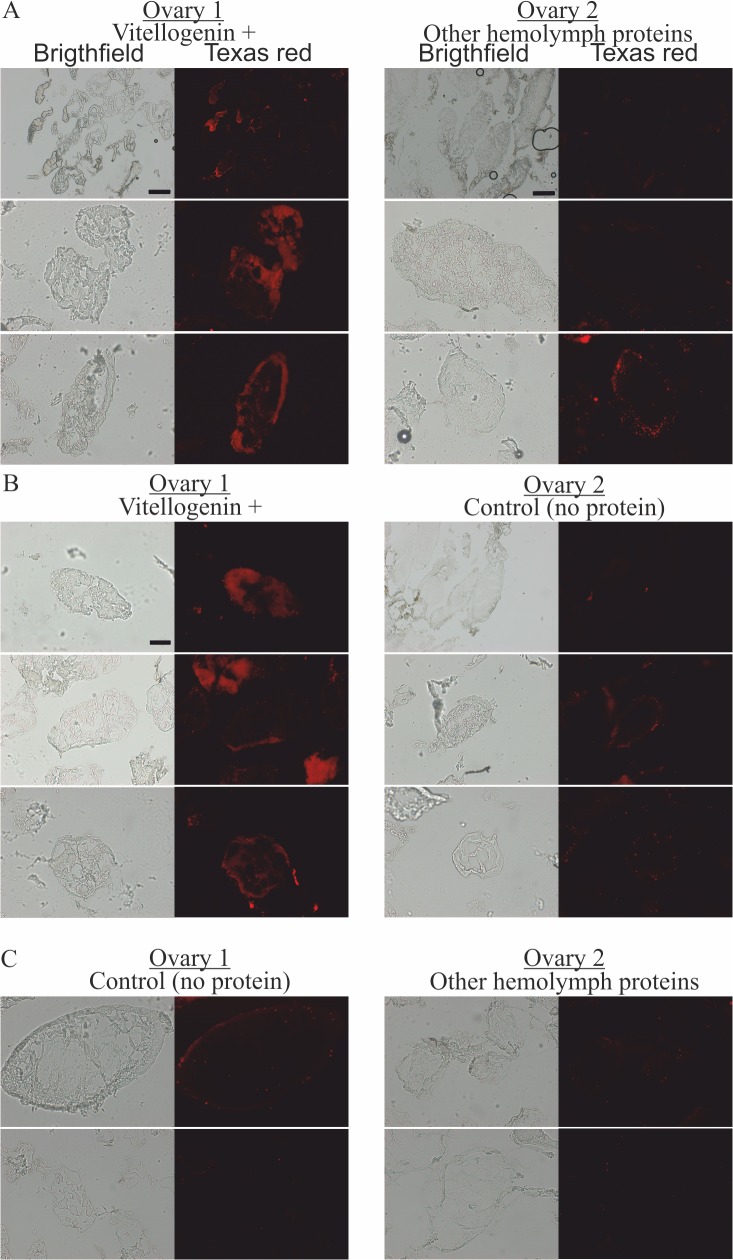
Fig 3. The localization of fluorescently-labeled bacterial fragments in cryo-sectioned honey bee queen ovaries incubated in the presence of pure Vg, in the presence of hemolymph proteins other than Vg, and in the absence of any externally provided protein.
(A) One ovary was incubated with Vg (left) and the other with other hemolymph proteins (right), N = 3. (B) One ovary was incubated with Vg (left) and the other without any protein (right), N = 3. (C) One ovary was incubated without any protein (left) and the other with other hemolymph proteins but Vg (right), N = 2. The scale is 50 μm, or 200 μm (the latter is indicated with scale bars).

