
Figure 2. Initiation of primordial germ cell migration.
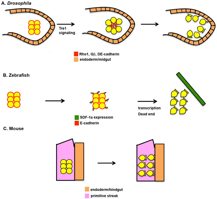
a | Drosophila melanogaster. I. At early stage 9 (∼4h After Egg Laying (AEL)), germ cells are tightly clustered in the midgut pocket. Primordial germ cells (PGCs) are not polarized at this stage and show little interaction with the midgut primordium. E-Cadherin, the small GTPase Rho1 and Gβ proteins are present uniformly at the cell periphery. Trapped in endoderm 1 (Tre1) signalling leads to the polarization of the PGCs, which take on a radial organization with the tails of the cells facing the inside of the cluster and the leading edges facing the midgut primordium. E-Cadherin, Rho1 and Gβ are redistributed to the tails of the cells. Next, the PGCs lose adhesion to each other and begin to extend cellular protrusions towards the epithelial cells of the midgut. b | Zebrafish. At specification, PGCs have a smooth, round morphology and do not posses migratory activity (3 hours post-fertilization (hpf)). PGCs begin to randomly extend small cellular protrusions in multiple directions at 3.5hpf. These protrusions disappear during mitosis. At 4.5hpf, PGCs become polarized, individualize and extend broad protrusions at the leading edge. This step is dependent on transcription and the Dead End protein, and is necessary for the cells to respond to stromal derived factor 1a (SDF-1a, also known as CXCL12a) chemokine signalling. c | Mouse. Following specification in the posterior primitive streak (embryonic day 7.5), PGCs have a smooth, round morphology. PGCs acquire a polarized morphology prior to initiating their migration into the endoderm. The molecular mechanisms regulating this polarization are not understood.