FIGURE 4.

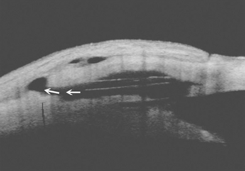
Anterior segment optical coherence tomography. The intrascleral lake and supraciliary space after the implantation with transcleral outflow (arrows) can be observed.

Anterior segment optical coherence tomography. The intrascleral lake and supraciliary space after the implantation with transcleral outflow (arrows) can be observed.