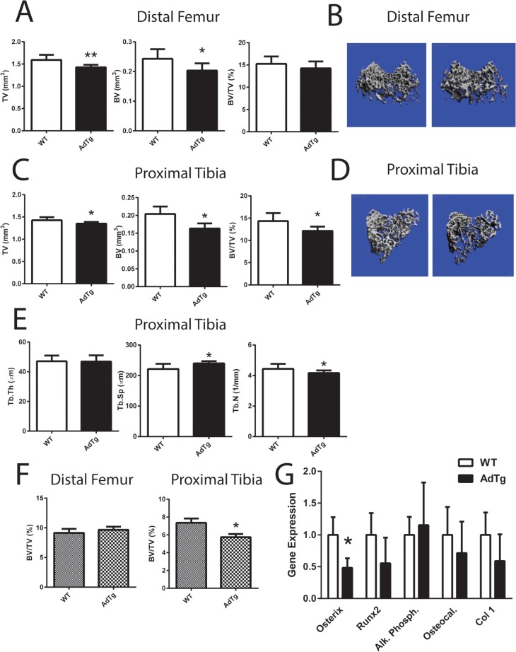
Fig 1. Cancellous bone measurements.
Bone tissue volume refers to the entire volume of bone scanned (TV), bone volume refers to the volume of mineralized bone within the scanned region (BV) and fractional bone volume referring to the fraction of mineralized bone relative to the total bone volume (BV/TV) was assessed by A) μCT and B) three-dimensional reconstruction μCT renderings at the distal femur and C) TV, BV, and BV/TV at the proximal tibia D) three-dimensional reconstruction images at the proximal tibia D). E) Trabecular thickness (Tb.Th), spacing (Tb.Sp.), and number (Tb.N) was assessed at the proximal tibia by μCT analysis. F) Histomorphometric analysis of distal femur and proximal tibia using Bioquant Software. G) Expression level of osbteoblast marker genes: Osterix, Runx2, Alkaline Phosphatase (Al. Phosph.), Osteocalcin (Osteocal.) and Collagen Type I (Col 1). All expression data were obtained by RT-qPCR analysis of RNA and calculated based on the ddCt method with GAPDH as the reference gene and WT as the control. All data are shown as mean ± SEM from 12 week old female mice (n = 5–8). Statistical significance * P<0.05, ** P<0.01, WT compared with AdTg bones.

