
Fig 5. A potential prokaryotic new finding.
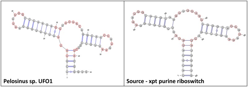
The predicted secondary structure drawing of an additional new bacterial aptamer domain detected by our method, in comparison to the known xpt guanine-binding aptamer domain. The aptamer sequence does not align via BLAST with the xpt riboswitch. Shown in red are preserved nucleotides.