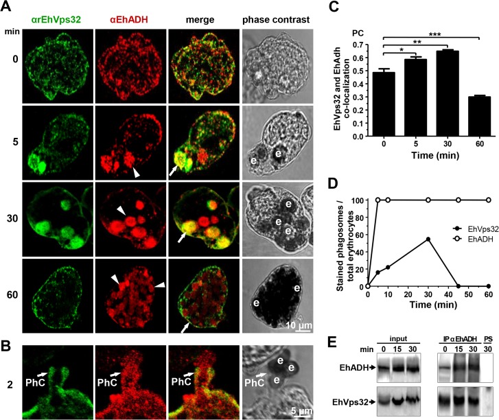
Fig 2. Co-localization and interaction of EhVps32 and EhADH during erythrophagocytosis.
(A,B) Trophozoites were incubated with erythrocytes for different times and analyzed through confocal microscopy. (A) After erythrophagocytosis, trophozoites were incubated with αrEhVps32 and αEhADH antibodies, then with FITC and TRITC-labeled secondary antibodies, respectively. (B) Illustration of a phagocytic cup (PhC). Arrows: co-localization of EhVps32 and EhADH. Arrowheads: EhADH in phagosomes without EhVps32 signal. e: erythrocytes. (C) Pearson’s coefficient (PC) to quantify EhVps32 and EhADH co-localization in the entire cell. (*) p<0.05, (**) p<0.01 and (***) p<0.001. (D) Proportion of erythrocytes inside phagosomes decorated by αrEhVps32 and αEhADH antibodies with relation to total ingested erythrocytes per trophozoite. (E) Immunoprecipitation of trophozoites lysates at different times of erythrophagocytosis using αEhADH antibodies or preimmune serum (PS).

