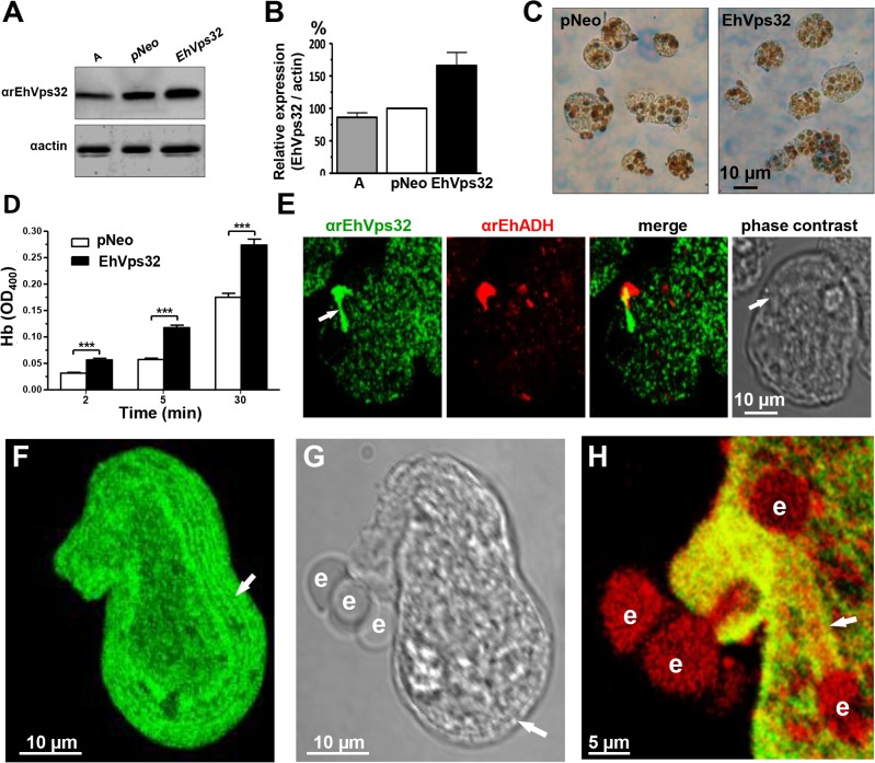
Fig 7. Expression and localization of EhVps32 and rate of erythrophagocytosis in pNeoEhvps32-HA transfected trophozoites.
(A) Western blot assays of trophozoites lysates from wild type clone A (lane A) and pNeo (lane pNeo) and pNeoEhvps32-HA (lane EhVps32) transfected cells, using the αrEhVps32 antibodies. As a loading control, the same membrane was re-blotted with αactin antibodies. (B) Densitometry analysis of bands showed in (A), normalized against actin band. (C) Diaminobenzidine-stained trophozoites that ingested erythrocytes for 30 min. (D) Rate of erythrophagocytosis of transfected trophozoites. (***) p<0.001. (E) Confocal microscopy of pNeoEhvps32-HA transfected trophozoites in resting conditions, using αrEhVps32 and αEhADH antibodies, followed by FITC-labeled and TRITC-labeled secondary antibodies. Arrows: membrane projections. (F-H) Trophozoites overexpressing EhVps32 incubated with erythrocytes for 2 min treated with αrEhVps32 and αEhADH antibodies and FITC- and TRITC-secondary antibodies, respectively. (F) Maximum projection of a transfected trophozoite evidencing the concentric arrays (arrow) formed by EhVps32. (G) Phase contrast. Arrow: concentric arrays. (H) Merging image (αrEhVps32 and αEhADH antibodies). Arrow: tunnel-like structure. e: erythrocytes.

