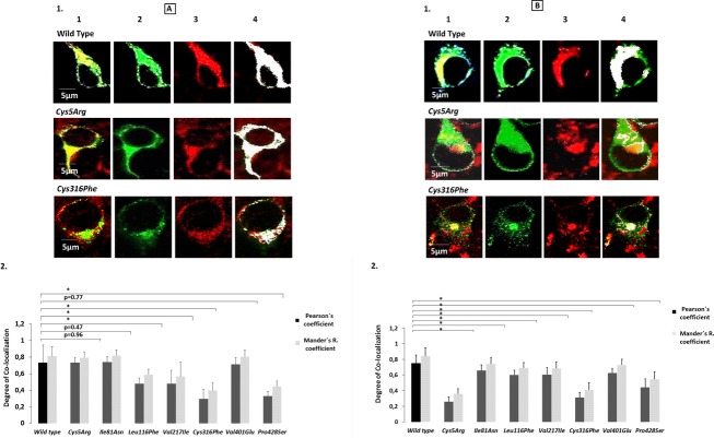
Figure 2.
Confocal analysis and the degree of colocalization for wild-type and the reported missense mutations. A(1) Confocal images for colocalization of our wild-type and mutant variant FXIIIB subunit protein with ER. In order to avoid any confusion only the two cysteine-based mutations, Cys5Arg and Cys316Phe have been shown here. The images for all the missense mutations can be seen in Figure S3. Each representative confocal image is split into four sections: green staining representing the FXIIIB subunit protein, red staining representing the ER, yellow staining showing the colocalization of ER and FXIIIB subunit and finally white dots which also represents co-localization of ER and FXIIIB subunit. A(2) Bar graph representing comparative degree of ER colocalization with ER for the wild-type and mutant variant FXIIIB subunit protein. B(1) and B(2) is similar to A(1) and A(2), the main difference being that the red stain represents the Golgi network and the co-localization overlays shown are for the FXIIIB subunit protein with the Golgi network.

