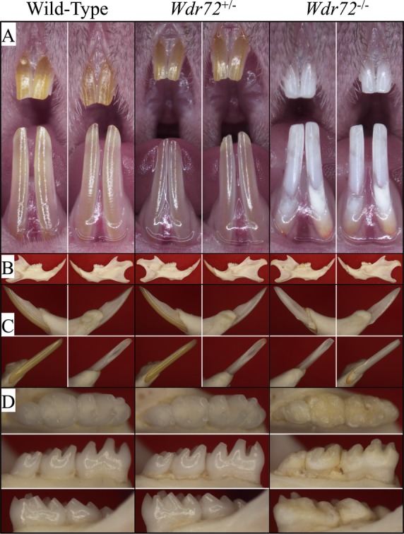
Figure 2.

Photographs of 7-week-old mice of three Wdr72 genotypes. (A) Frontal view of maxillary and mandibular incisors. The null incisors exhibited a chalky-white appearance and chipped incisal edges. (B) Medial (left) and lateral (right) views of hemimandibles. The size and the bony structures of the mandible in null mice appeared normal and comparable to those of the wild-type and heterozygous mice. (C) Lateral (upper left), medial (upper right), labial (lower left), and lingual (lower right) views of mandibular incisors (D) Occlusal (upper), lingual (middle), and buccal (lower) views of mandibular molars. The Wdr72−/− molars showed extensive attrition with dull, yellowish areas of exposed dentin.
