
Figure 4.
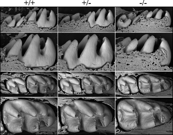
Backscattered SEMs of Day 14 Wdr72 mouse mandibular molars following soft tissue removal. The top two rows are lingual views; the bottom two rows are occlusal views. The sizes of the teeth and the thicknesses of the enamel appeared to be similar in the three genotypes. These images show the molars prior to eruption. Immediately after eruption the enamel layer is abraded away.