
Figure 5.
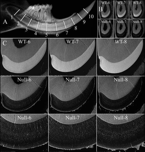
Backscattered SEMs of 7-week mandibular incisor cross-sections from late maturation stage. (A) Hemimandible radiograph showing the 1 mm-incremental cross-section levels from apical to incisal along a mandibular incisor. (B) Low-magnification views of mandibular cross-sections at levels 6–8, of wild-type (top) and null (bottom) incisors. (C) Higher magnification bSEM images of level 6–8 cross-sections. The electron densities of the Wdr72−/− enamel at all three levels were similar to each other and significantly lower than those of the wild-type enamel (upper panel), demonstrating a severe hypomineralization defect of the Wdr72−/− enamel. Also, an irregular layer of electron-dense crust covered the enamel of the null mice. The complete panel of cross-sections (levels 2–8) for the three genotypes is presented in Fig. S6. bSEM, backscattered scanning electron microscopy.