Figure 7.

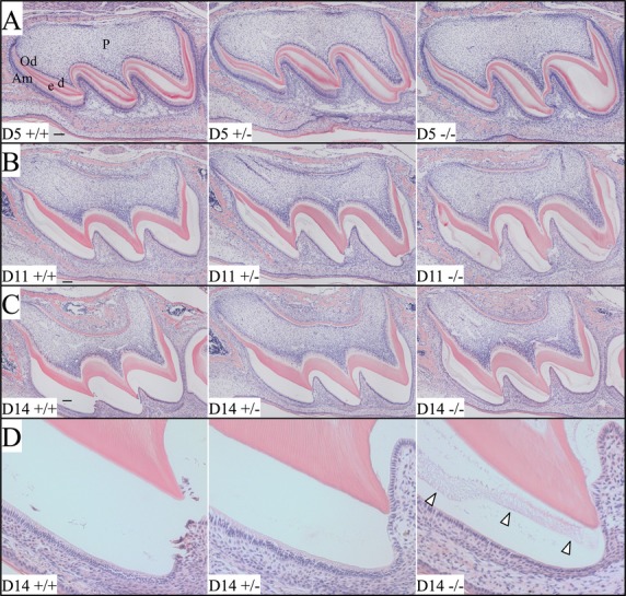
Histology of D5, D11, and D14 maxillary first molars. (A–C) Low-magnification (10×) views of the D5, D11, and D14 maxillary first molars. (D) Higher magnification (20×) views of the D14 first molar mesial cusp detailing residual matrix (arrowheads) in the null molar. At D5, enamel matrices are actively being secreted by ameloblasts and show eosinophilic staining. No evident histological differences could be identified between the first molars of the three genotypes at D5. At D11, reabsorption of extracellular matrix protein was most advanced in the wild-type and heterozygous mice. Residual protein was greatly diminished near the cusp tips where enamel maturation first commenced. By D14 (immediately prior to eruption), most of the protein in the extracellular matrices had been reabsorbed in the wild-type and heterozygous molars, while the null molar retained eosinophilic material in the enamel space (arrowheads). Am, ameloblasts; d, dentin; e, enamel; Od, odontoblasts; P, dental pulp. Scale bars: 100 μm.
