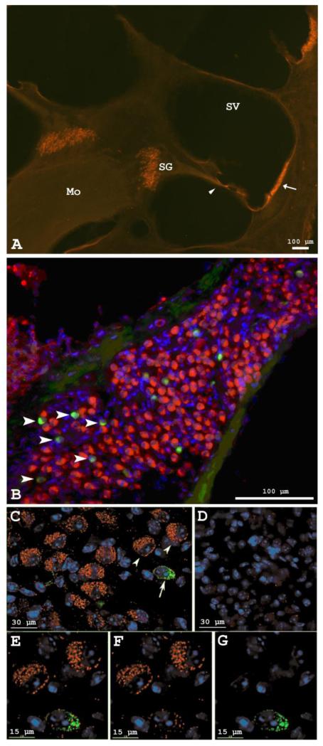
Fig. 1.
Neuroglobin protein distribution in the cochlea. A Ngb-immunofluorescence (red) in spiral ganglion neurons, stria vascularis (arrow) and basilar membrane (arrowhead) of the rat cochlea. Mo=modiolus; SV=scala vestibuli; SG=spiral ganglion cells in Rosenthal’s canal. Scale bar=100 μm. B Double-label immunostaining of mouse spiral ganglion cells with anti-Ngb (red) and anti-peripherin (green). SgnI cells were immunolabeled with anti-Ngb while SgnII cells (arrowheads) were immunostained with anti-peripherin. Nuclei are stained blue with bisbenzimide. C-G Deconvolution images illustrating differential Ngb immunostaining in SgnI and SgnII cells of the mouse cochlea. C Double-label immunostaining with anti-Ngb (red: arrowheads) and anti-peripherin (green: arrow). D Control omitting primary anti-Ngb and anti-peripherin antibodies. E-G High-power images of the three cells indicated by arrows and arrowheads in C: E SgnI and SgnII (combined red and green channels), F SgnI only (red without green channel), and G SgnII only (green without red channel). Nuclei are stained blue with bisbenzimide.

