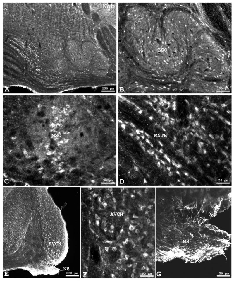
Fig. 2.
Neuroglobin (Ngb) protein distribution in the rat superior olivary complex and cochlear nucleus of the rat. A Ngb-immunofluorescence in different subnuclei of the SOC in the right half of the brainstem. Higher magnifications are given in B of the lateral superior olive (LSO), in C of the medial superior olive (MSO) and in D of the medial nucleus of the trapezoid body (MNTB). Orientation in each section is: medial left, dorsal up. E Neuroglobin protein distribution in the anteroventral cochlear nucleus (AVCN) and cochlear nerve. Higher magnfications demonstrating positive neuronal perikarya are given in F of the AVCN and in G of the Ngb-immunoractive fibers in the cochlear root of the olivocochlear nerve (N8), shown in extended focal imaging. Orientation in each section is: medial left, dorsal up.

