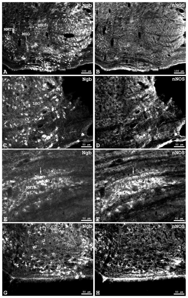
Fig. 6.
Double immunofluorescence labelling of neuroglobin (Ngb, shown in A,C,E,G) and neuronal nitric oxide synthase (nNOS, shown in B,D,E,H) in frontal sections of the mouse superior olivary complex. A,B: overview. C,D: higher magnification of double-labelled neurons (depicted by “1”) in the lateral superior olive (LSO). Neurons showing only Ngb-immunoreactivity were also found (“2”). Middle is left, dorsal is up. Higher magnifications from mouse medial nucleus of the trapezoid body (MNTB) are shown in E,F where a group of double-labelled neurons is marked by arrows, and in the ventral periolivary region (G,H) where double-labelled neurons are depicted by “1” and those showing only Ngb-immunoreactivity by “2”.

