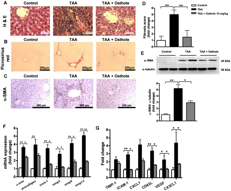
Fig. 2.

Osthole attenuated liver injury and fibrogenesis in the TAA rat model. Representative liver sections were obtained from groups as follows: control, TAA and osthole-treated TAA rats. Liver sections were analyzed with a H&E-, b Picrosirius red- and c immunostaining of α-SMA. d Fibrosis scores were assessed by a pathologist in a blind fashion. e Quantification of collagen-positive area was performed using Metamorph software. f α-SMA protein in liver tissues detected by Western blotting analysis. g The expressions of α-sma and procollagen I, icam-1, mmp2, mmp9 and mmp13 transcripts in livers measured by qRT-PCR. h ELISA of TIMP-1, ICAM-1, CXCL7, CD62L, VEGF, and CX3CL1 in plasma of various groups. Data are shown as mean ± SD of 8 rats in each group. *p < 0.05; **p < 0.01, compared with other groups
