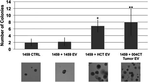
Fig. 1.

Extracellular vesicle-mediated induction of soft-agar growth. EVs were isolated from malignant (HCT116) cells and from patient tumor tissue (004CT Tumor). 1459 cells were co-cultured for 7 days with HCT116 EVs and 004CT Tumor EVs. In both experiments, cells were harvested and utilized for soft agar assay. 1459 cells were also co-cultured with EVs isolated from 1459 cells. Soft agar cloning was performed for 2 weeks and cell colonies were counted with 5 fields/dish using the 40× objective. There were 5 dishes/condition. To provide an estimation of colony size that was evaluated, the area for the colonies counted in an average field was determined. The average area (μm2) determined were: 1459 CTRL, 1988; 1459 + 1459EV, 2603; 1459 + HCT EV, 1860; 1459 + 004CT Tumor EV, 3841. The data represents the mean +/− s.d. of 2 independent experiments performed in triplicate. A paired t-test was performed to analyze the increase in soft agar colony formation of 1459 + HCT116 EVs when compared to untreated 1459 cells, *p < 0.00001. Increase in colony formation of 1459 + 004CT Tumor EV compared to untreated 1459 cells, **p < 0.00001, was also assessed
