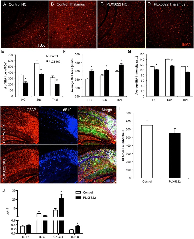
Fig. 5.

Lower-dose CSF1R inhibition partially reduces microglia numbers. The brains of 3 months 3xTg-AD-treated mice were examined for effects of PLX5622 on pathology. a–d IBA1 immunofluorescent staining was performed and representative 10× images are shown of control and treated hippocampus and thalamus. e IBA1+ cell counts revealed a reduction by 30 % in the treated groups. f, g IBA1+ cells in treated brains are larger but have reduced staining intensity as compared to 3xTg-AD untreated mice. h Immunofluorescent staining for the astrocytic markers GFAP (red), S100 (green), and plaques with 6E10 (blue), with the hippocampal region shown. i Quantification of the number of GFAP+ cells in the hippocampal sub-field. j Inflammatory profiling of whole brain homogenates shows significant increases in CXCL1 and TNFα but not Il-1β or Il-6. *Indicates significance (p < 0.05) by unpaired Students t test. Error bars indicate SEM
