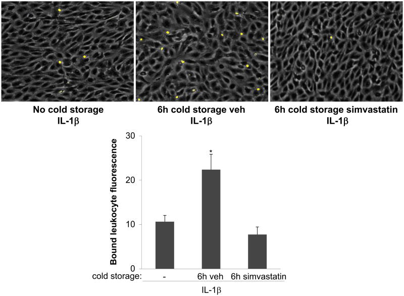
Figure 4. Simvastatin inhibits endothelial activation after cold storage.
Top, EC monolayers were cultured for 24h under vasoprotective flow, followed by the immediate addition of IL-1β 0.1 U/mL, 4h, 37°C, left), or by cold storage for 6h with simvastatin (1 μM, right) or its vehicle (center), and then treated for 4h with IL-1β (as above). All groups were then incubated with fluorescently labeled HL-60 cells. Shown are representative fields of EC monolayers (gray) and attached HL60 cells (yellow). Bottom, quantitative fluorescence analysis of bound HL-60 cells (*p<0.05 vs. no cold storage and 6h simvastatin, n=4).

