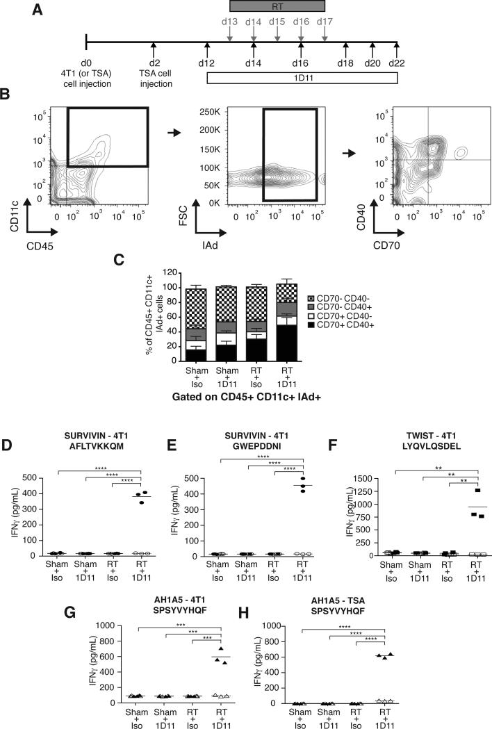
Figure 1. TGFβ blockade with RT enhances TIDC activation and induces CD8+ T cells responses to endogenous tumor antigens.
(A) Treatment schema. (B-C) Analysis of 4T1 TIDC at day 22 (n=9/group). To obtain sufficient material, 3 tumors were pooled to obtain 3 independent samples for each group. Viable cells were gated on CD45+CD11c+IAd+ TIDC and analyzed for expression of activation markers CD40 and CD70. (B) Representative dot plots. (C) Bar graphs showing significant increase in mean percentage of TIDC expressing CD40 and CD70 in tumors of mice treated with RT+1D11 (P<0.005 compared to all other groups). (D-H) IFNγ production by CD8+ T cells from LN draining 4T1 (D-G) or TSA (H) tumors in response to peptides derived from survivin (D and E, closed circles), Twist (F, closed squares,), and gp70 (AH1A5) (G and H, closed triangles) or irrelevant peptide (open symbols). Each symbol represents one animal. Horizontal lines indicate the mean of antigen-specific (solid lines) or control (dashed lines) IFNγ concentration. Data are representative of three independent experiments. **p<0.005; ***p<0.0005; ****p<0.00005.

