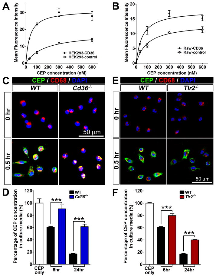
Figure 5. CEP binding and scavenging by CD36 and TLR2.
A and B. Cell-based CEP binding assay using HEK293 (A) and Raw264.7 (B), which were stably expressing CD36. CEP binding to the cells was visualized by FACS after staining the cells with rabbit anti-CEP antibody and Alexa-488-conjugated anti-rabbit IgG secondary antibody. MFI are shown. C. Reduced CEP binding to CD36 null cells. Immunofluorescence of CEP, CD68 and DAPI on the primary WT and Cd36−/− R-MPMs, which were incubated at 37 °C with 500 nM CEP-BSA for 30 min. D. Reduced CEP scavenging by CD36 null cells. WT and Cd36−/− R-MPMs were treated with 500 nM CEP-BSA for 24 hours at 37 °C, then the supernatants were collected. CEP levels were measured by ELISA and residual CEP values are shown. The amount of CEP without added R-MPMs was assigned a value of 100%. E. Reduced CEP binding to TLR2 null cells. Immunofluorescence of CEP, CD68 and DAPI on the primary WT and Tlr2−/− R-MPMs, which were incubated at 37 °C with 500 nM CEP-BSA for 30 min. F. CEP scavenging by Tlr2−/− cells is reduced. WT and WT and Cd36−/− R-MPMs were treated with 500 nM CEP-BSA for 24 hours at 37 °C, then the supernatants were collected. CEP levels, measured by ELISA are shown. Amount of CEP without added R-MPMs was assigned a value of 100%.

