
Fig. 5.
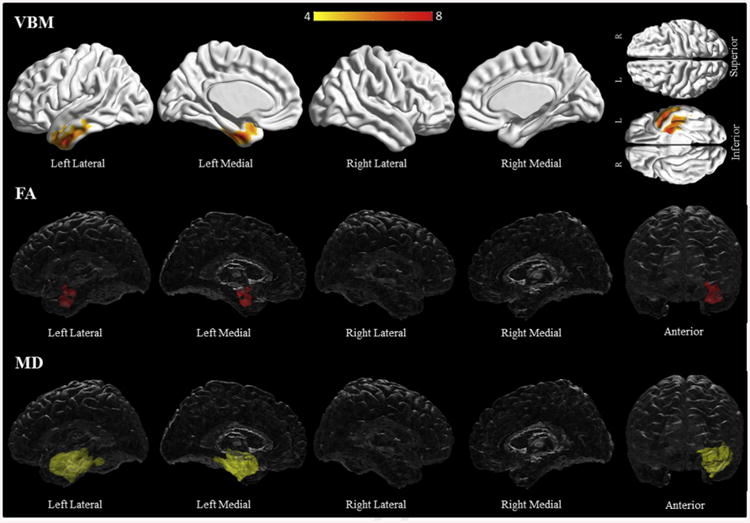
Results of the voxel-based morphometry and diffusion tensor imaging analysis in the progressive fluent aphasia group. Patterns of grey matter loss on voxel-based morphometry (top), and reduced fractional anisotropy (middle) and increased mean diffusivity (bottom) on diffusion tensor imaging analysis, in the progressive fluent aphasia group compared with controls. All results shown at p < .05 (corrected for multiple comparisons using family-wise error). Results are shown in 3D renderings of the brain.