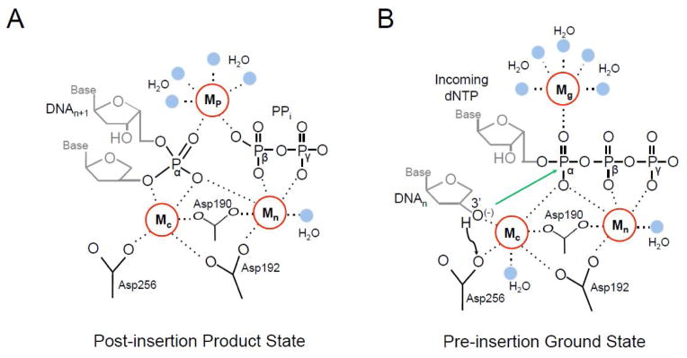
Fig. 4.

Adjunct metal binding sites in the pol β active site. (A) The post-insertion product state of the pol β active site is shown following insertion of the incoming dNTP (DNAn+1). The catalytic (Mc), nucleotide (Mn), and product (Mp) metal binding sites are shown as red circles with dashed lines to coordinating groups. Four Mp coordinating water molecules are in blue. Active site aspartic acid residues, the remaining bound pyrophosphate (PPi) group, and a coordinating water molecule (blue) are indicated. The groups corresponding to Pα, Pβ, and Pγ of the inserted dNTP triphosphate are labeled for clarity. The Mc binding site has only five coordinating ligands due to the missing 3′-OH. This results in the catalytic Mg2+ vacating the active site and being replaced by a Na2+ for pol β. (B) The pre-insertion state of the pol β active site is shown with an incoming dNTP and a primer terminus with a 3′-OH (i.e., 2′-deoxyribose). The black arrow corresponds to deprotonation of the 3′-OH to Asp256. The green arrow indicates the resulting O3′ anion attack on Pα. The catalytic (Mc), nucleotide (Mn), and ground (Mg) metal binding sites are shown as red circles with dashed lines to coordinating groups. Active site aspartic acid residues and coordinating waters (blue) are shown.
