
Figure 1.
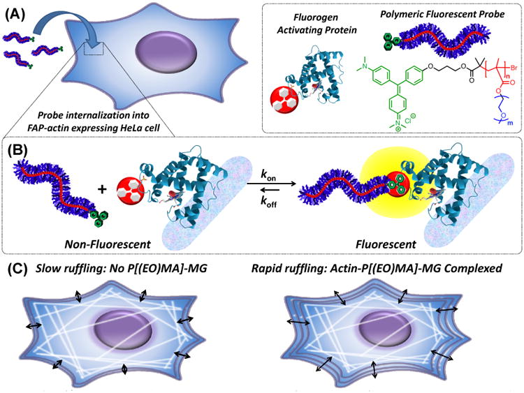
(A) Illustration depicting internalization of a polymer fluorogen into a genetically targetable actin-modified HeLa cell. Legend and schematic breakdown of the polymer fluorogen consisting of MG (green), methacrylate backbone (red) with a DP of “n” repeat units, and ethylene oxide (EO) side-chains (blue) with “m” repeat units. Subsequent data in Figure 3 and 4 are color-coded for clarity, where red figures indicated backbone DP variations and blue EO side-chain variations. (B) Polymeric fluorescent probe disassociated and complexed with a FAP representing its non-fluorescent and fluorescent states, respectively. (C) Illustration of binding polymer fluorogens to FAP-fused actin and their effect on cellular ruffling behavior.