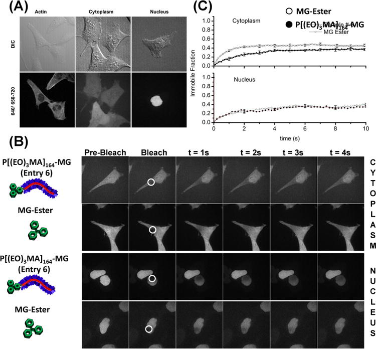
Figure 5.

Live-cell confocal microscopy images of cells stably expressing targeted FAP proteins. (A) FAP-Actin (HeLa), Cytoplasm (HEK-293), and Nuclear targeted proteins all show proper localization when labeled with the polymeric fluorogen P[(EO)3MA]164-MG (Table 1, Entry 6). Cells were incubated in DMEM with a 500 nM polymeric dye for 12 h. Emission of MG bound to FAPs was observed between 650-710 nm and excited with a 633 nm laser. Additional Actin images are supplied in the supporting information (Figure S5). (B) Fluorescence recovery after photobleaching (FRAP) images for polymeric dye and MG ester in the cytoplasm and nucleus. The cells were imaged for a single frame at an exposure of a 100 ms followed by bleaching in the region shown using a circle with a white border. The subsequent images show the recovery of the signal in a 4s duration. (C) Plots of average intensity as a function of time in the circular regions shown in B, in nucleus and cytoplasm for polymeric dye and MG ester. Scalebar in confocal images is 20 μm.
