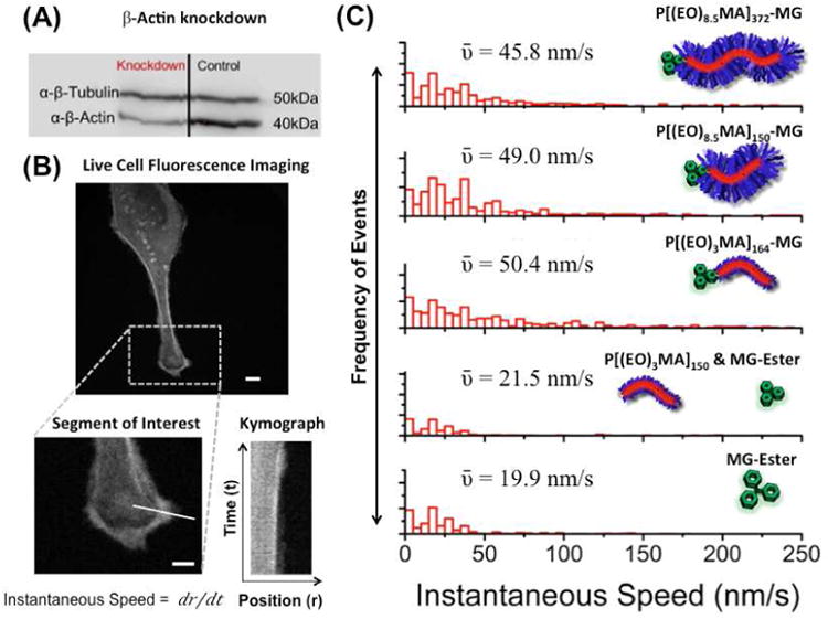
Figure 6.

Effect of actin bound polymer fluorogen on membrane ruffling. (A). Western blot showing the knockdown of β-actin by siRNA in HeLa cells expressing modified actin. (B) Fluorescence images of HeLa cells after knockdown and incubation with 500 nM polymeric dye or MG ester in DMEM + 10% FBS for 6 hours. A segment of interest was chosen along the cell membrane and a kymograph was generated for the selected segment. Scalebars in confocal images represents 10 μm. From the kymograph, instantaneous speeds were obtained at selected segment for various time points. (C) Histograms of the instantaneous speed are shown for the knockdown cells with various polymer fluorogens and control systems with their corresponding mean-instantaneous speed values (ῡ).
