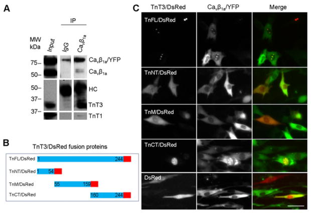
Figure 3. Interaction between Cavβ1a and TnT3 in C2C12 cells.
(A) C2C12 cells transduced with RAdCavβ1a/YFP were cultured until myotubes formed. Whole cell lysis preparations were immunoprecipitated with Cavβ1a antibody or IgG and immunoblotted. The antibody against heavy chain (HC) indicates a similar Cavβ1a antibody and IgG loading. (B) Diagram of the DsRed-conjugated TnT3 constructs. (C) Co-expression of Cavβ1a/YFP and various TnT3/DsRed constructs in C2C12 cells cultured in growth medium. Both TnFL/DsRed and TnCT/DsRed localized in the nucleus and co-localized with Cavβ1a/YFP. In contrast, Cavβ1a/YFP expression, alone or co-expressed with TnNT/DsRed, TnM/DsRed, or control DsRed, was localized throughout the cell. Scale bar = 50 μm.

