. 2015 Jul 28;9(4):565–572. doi: 10.4184/asj.2015.9.4.565

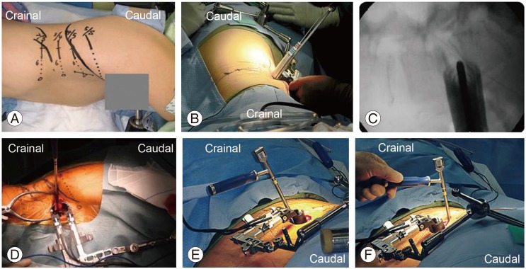
Fig. 2. (A) Skin marking to check the disk level using a C-arm X-ray imager. The skin incision was made 6 to 10 cm anterior to the midportion of the disk. A longitudinal incision from 3 to 4 cm is recommended. The surgeon cleared the peritoneum from the psoas using a finger. (B) Sequential dilation was used. (C) C-arm X-ray image after dilation. (D) Retractor for oblique lateral interbody fusion was used after dilatation. A trial cage was used after removal of the intervertebral disk. (E, F) A Clydesdale Spinal System cage filled with autologous bone is implanted. Implantation starts from a lateral oblique direction; finally the cage is inserted from a true lateral direction.

Fig. 2