. 2015 Jul 28;9(4):636–644. doi: 10.4184/asj.2015.9.4.636

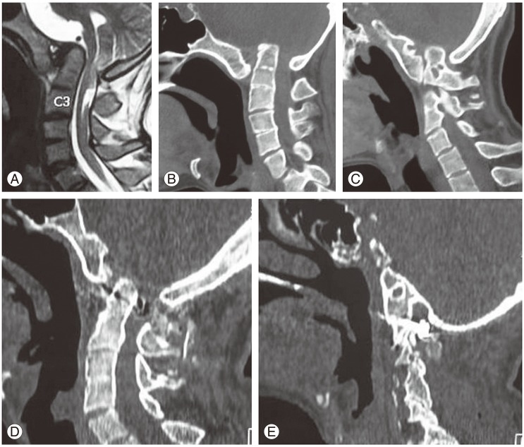
Fig. 2. Images of a 32-year-old male. (A) T2-weighted magnetic resonance imaging shows group A basilar invagination. Chiari malformation is observed. Ill-defined cord changes are observed in the spinal cord at C3 spinal level. (B) Computed tomography (CT) scan showing basilar invagination. Assimilation of atlas and C2-3 fusion can be observed. (C) Sagittal image with the section passing through the facets. Facets of C1 and C2 are in severe mal-alignment. (D) Postoperative image following surgery that involved distraction, reduction and fixation of the atlantoaxial joint. Please note the reduction of basilar invagination. (E) CT scan image showing realignment of the facets.

Fig. 2