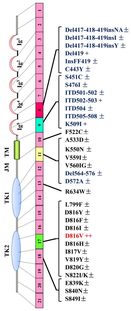
Figure 2. Representation of the structure of KIT, illustrating the localization of the more frequently observed mutations in the KIT sequence in pediatric and adult patients with mastocytosis.
The receptor is presented under its monomeric form, whereas its wild-type counterpart dimerizes upon ligation with SCF before being activated in normal cells. In children,he KIT D816V PTD mutant (in red) is found in nearly 30% of the patients, whereas the ECD mutants (in blue) are found in nearly 40% of the affected children. In adults, depending of the categoty of mastocytosis, the KIT D81V mutant is found in at least 80% of all patients. The complete list of KIT mutants retrieved in the literature for mastocytosis is depicted here. In children, the structure of KIT is found WT in around 25% of the patients analyzed, whereas in adults, KIT is found WT in less than 20% of all patients analyzed so far. Some of the mutations (in black) are found only in a very few number of patients. Del: deletion; ECD: Extracellular domain; Ins: Insertion; ITD: Internal tandem duplication; JMD: Juxtamembrane domain; KI: Kinase insert; PTD: phosphotransferase domain; TMD: Transmembrane domain. ±: mutation found in less than 1% of the patients; +: mutation found in 1 to 5% of the patients; ++: mutation found in around 30% of pediatric patients and in > 80% of all adult patients.

