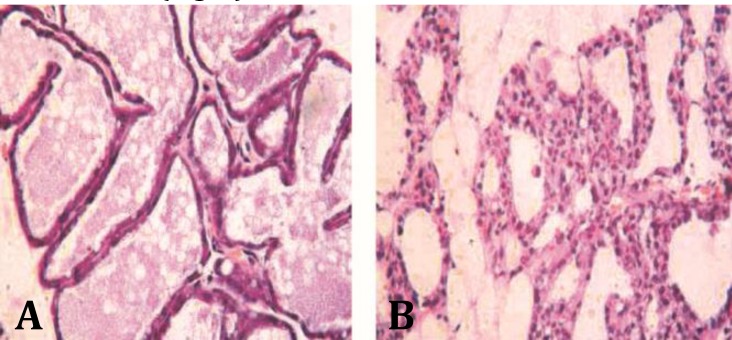
Fig. 1.
Histopathology of rat mammary gland at 12 hr after intra-mammary infusion LPS, (A) control group: Mammary gland tissue, epithelial tight junction, intact acinar structure and no infiltration of inflammatory cells in mammary gland, (B) LPS infused group: inflammatory cells were infiltrated in mammary tissue, acinar structure and acinar lumina were also disrupted

