
Figure 9.
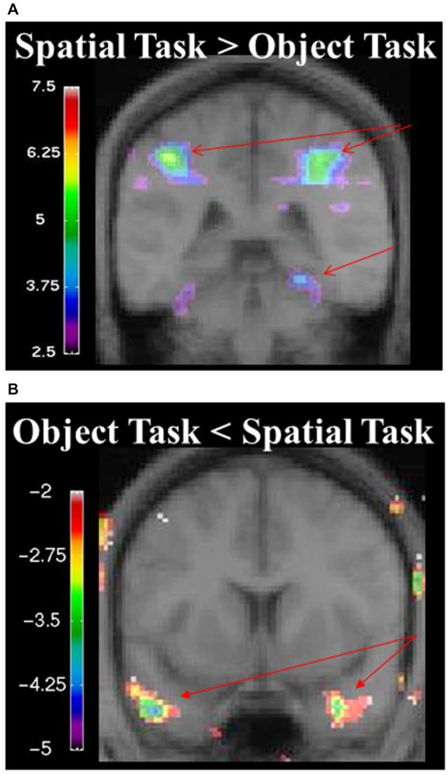
(A) fMRI results from the spatial memory configural task contrasted against the object identity task. Results are overlaid on the mean structural image. Hemispheres are indicated by L (left) and R (right). Increase in activity in the RPH (x = 24, y = −38, z = −16; t = 3.79, p < 0.005), and bilateral activation in the parietal lobes (left: x = −26, y = −56, z = 42; t = 8.32, p < 0.00005 right: x = 26, y = −60, z = 36; t = 8.64, p < 0.00005). (B) fMRI results from the object identity task contrasted against the spatial memory configural task. Significant increase activity in the bilateral anterior temporal lobe (left: x = −46, y = 8, z = −34; t = −4.15, p < 0.005; right: x = 34, y = 0, z = −28; t = −3.99, p < 0.005). The negative values in the object identity task represent greater activity during that task when it was contrasted to the spatial task.