Figure 5.
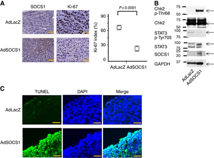
SOCS1 showed antitumour activity in a GC xenograft model: histological analysis of peritoneally disseminated tumours after SOCS1 gene therapy. (A) Immunohistochemical analysis of SOCS1 and Ki-67 in MKN45-Luc tissue from animals treated with AdSOCS-1 or AdLacZ. Scale bar=20 μm. AdSOCS1 group showed increased SOCS1 expression in tumour and significantly decreased Ki-67 index. Ki-67 staining was recorded as the ratio of positively stained cells to all tumour cells in 10 fields ( × 200 magnification). Statistical analyses were performed using Welch's t-test and two-sided P values less than 0.05 were considered significant. Values shown represent average±s.d. (B) The peritoneally disseminated tumours treated with Ad-SOCS1 were analysed by western blotting. We confirmed the expression of SOCS1 and decreased levels of pSTAT3. Phosphorylation of Chk2 at Thr68 was also increased, the same as in vitro. (C) Immunohistochemical analysis by TUNEL (blue fluorescence, DAPI staining for nuclei; cyan fluorescence, TUNEL positive) in MKN45-Luc tissues from animals treated with AdSOCS1 or AdLacZ. Scale bar=20 μm.

