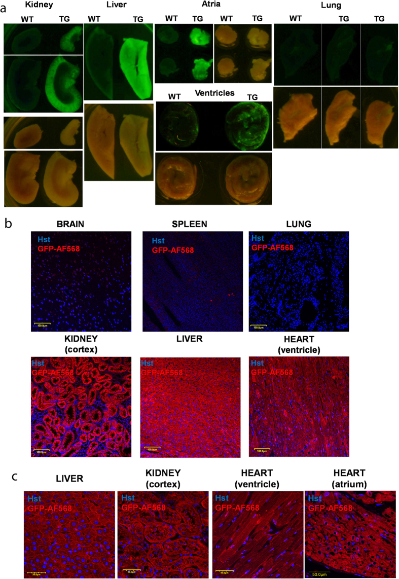
Figure 2. Expression of the GCaMP2 calcium sensor in transgenic rats.
(A) Fluorescence (upper) and bright field (lower) images of wild type (WT) and transgenic (TG) kidney, liver, heart atria, ventricles and lung. (B) Immunohistochemistry of transgenic brain, spleen, lung, renal cortex, liver, and heart ventricle at lower magnification (scale bars represent 100 μm). (C) Immunohistochemistry of transgenic liver, renal cortex, heart ventricle and atrium at higher magnification (scale bars represent 50 μm). Immunostaining of the GCaMP2 was performed by a GFP-recognizing antibody. Hoechst (Hst) was used to stain the nuclei.

