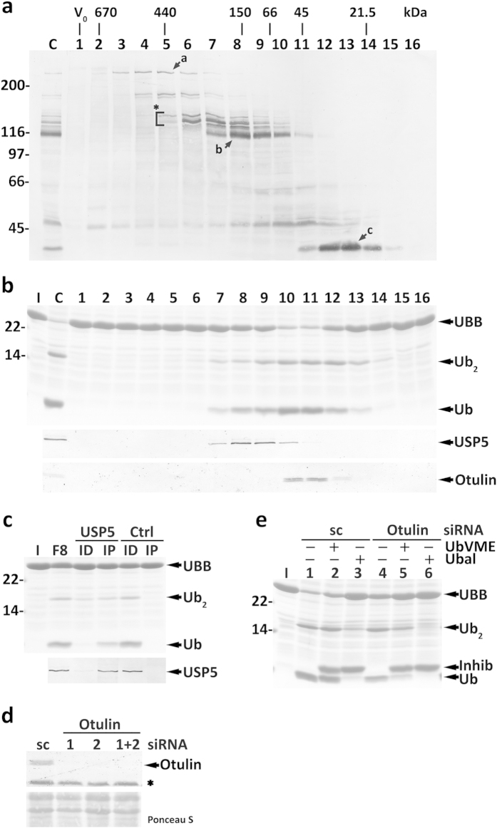
Figure 5. Characterization of HeLa cells cytosolic DUBs acting on human UBB.
(a) Total cytosolic proteins (lane C) and the corresponding SEC fractions (lanes 1–16) were reacted with HA-UbVME and analyzed by SDS-PAGE/western blot with an anti-HA antibody. The asterisk and arrows a-c indicate HA-UbVME-reactive DUBs that co-elute with enzyme activities described in the main text. The elution positions of molecular mass protein standards, as well as the void volume (V0), are indicated. (b) SDS-PAGE/Coomassie blue staining analysis of UBB processing by SEC fractions (upper panel). Lane C, total cytosolic proteins were also assayed. The elution profiles of USP5 (middle panel) and Otulin (lower panel) are also shown. (c) Fraction 8 from SEC was subjected to an immunoprecipitation/immunodepletion assay using control (lanes Ctrl) or anti-USP5 IgGs (lanes USP5). Fraction 8 (F8) and the corresponding immunoprecipitated (lanes IP) and immunodepleted fractions (lanes ID) were assayed for UBB processing activity (upper panel). The distribution of USP5 in the samples is shown (lower panel). (d) Otulin knockdown in HeLa cells. Western blot analysis of Otulin in HeLa cells transfected with Otulin-specific siRNA oligos #1 and/or #2, or with a scrambled (sc) control. The asterisk indicates a protein recognized by the anti-Otulin antibody in total homogenates, but not in cytosolic fractions. The corresponding Ponceau S-stained membrane is shown to assess protein loadings. (e) OTULIN knocked-down HeLa cell total extracts have decreased HA-UbVME-insensitive UBB processing activity. Inhib, HA-UbVME or HA-Ubal. In (b,c,e) the cleavage intermediate (Ub2), and ubiquitin (Ub) are indicated. Lanes I, recombinant UBB used in the assays. In (a–c,e) numbers to the left indicate the molecular weights of protein standards in kDa.

