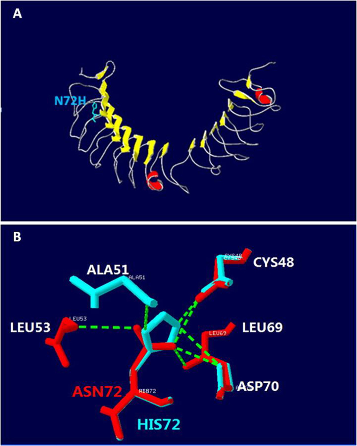
Figure 5. Structural model of nyctalopin built by the Swiss model.

(A) 3-Dimensional structure of nyctalopin. Yellow arrows show beta-strands. Red parts show α-helix. The side chains of N72 are marked in blue. (B) Detail of the effect of the N72H mutation showing loss of hydrogen bonds. The normal and mutated nyctalopin structures are shown in red and blue, respectively. Hydrogen bonds are shown by green dashed solid lines.
