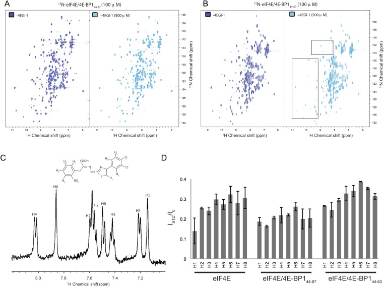
Fig. S8.
eIF4E/m7GTP/4E-BP1 complex with 4EGI-1. (A) 2D [1H, 15N] TROSY-HSQC correlated spectrum of 15N-eIF4E/m7GTP bound to 4E-BP144–87 or to (B) 4E-BP144–63, in absence (blue) or presence (cyan) of 4EGI-1. Slight overall chemical shift perturbations can be observed for 15N-eIF4E/m7GTP bound to 4E-BP144–87 upon addition of 4EGI-1. The peaks that are boxed in B are those for which intensities are severely reduced upon titration of 4EGI-1. (C) 1H-NMR spectrum of 4EGI-1 compound showing peak assignment for the aromatic region. 4EGI-1 molecular structure is shown in the upper part of the panel. (D) Saturation-transfer difference (STD) experiments, where magnetization is transferred form eIF4E alone or eIF4E complexed to 4E-BP144–87 or to 4E-BP144–63 to 4EGI-1. ISTD/I0 ratios for each 4EGI-1 aromatic ring proton in the presence of the different protein complexes are shown.

