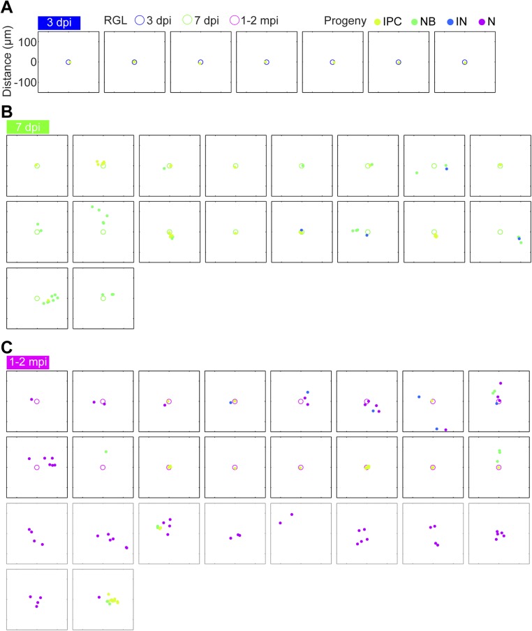
Fig. S1.
Tangential distribution among newborn neural progeny at 3 dpi, 7 dpi, and 1–2 mpi in the adult dentate gyrus. Two-dimensional SGZ plane projection of all RGL-containing clones containing at least IPCs at (A) 3 dpi, (B) 7 dpi, and (C) 1–2 mpi, plotted in a 300-μm-square window. RGLs are represented as open circles; neural progeny are represented as closed circles. Different time points or developmental stages are encoded by color. Clones that lacked RGLs at 1–2 mpi are also plotted in gray boxes and included in the histogram in Fig. 2C.

