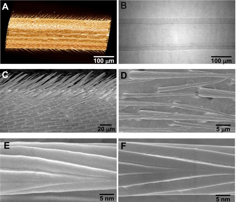
Fig. S1.
Microstructure of water strider leg. (A and B) Micro-XCT images indicate that the surface of water strider leg is composed of setae array, which is oriented toward the tip of the leg with ∼27° tilt angle. (C and D) SEM images of water strider leg reveal that the setae are all conical with an apex angle of ∼5°, in a periodicity of 5–10 μm between the neighboring setae. (E and F) Magnified SEM images show that the setae are composite of nanogrooves in longitudinal or quasi-helix orientation.

