
Fig. 6.
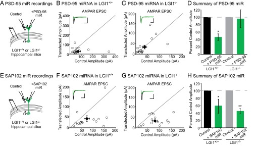
LGI1 is essential for PSD-95, but not SAP102, function. (A) Dual recordings of control and PSD-95 miRNA-expressing cells in LGI1+/+ or LGI1−/− slices. (B) Scatterplot of AMPAR EPSC amplitudes recorded from wild-type and PSD-95 miRNA-transfected cells. Representative traces of dual recordings are shown as Insets. (Scale bars, 50 ms and 50 pA.) (C) Scatterplot of AMPAR EPSC amplitudes recorded from LGI1−/− control and LGI1−/− cells transfected with PSD-95 miRNA. (Scale bars, 50 ms and 25 pA.) (D) Summary bar graphs showing normalized mean ± SEM of experiments in B and C. Reducing PSD-95 expression in wild-type slice culture results in a 50% reduction in AMPAR-mediated transmission (P = 0.03, n = 17). In LGI1−/− slices, reduction in PSD-95 expression has no effect on AMPAR-mediated transmission (P = 1.00, n = 16). (E) Dual recordings of control and SAP102 miRNA-expressing cells in LGI1+/+ or LGI1−/−. (F) Scatterplot of AMPAR EPSC amplitudes recorded from wild-type and SAP102 miRNA-transfected cells. Representative traces of dual recordings are shown as Insets. (Scale bars, 50 ms and 50 pA.) (G) Scatterplot of AMPAR EPSC amplitudes recorded from LGI1−/− control and LGI1−/− cells transfected with SAP102 miRNA. (Scale bars, 50 ms and 50 pA.) (H) Summary bar graphs showing the normalized mean ± SEM of experiments in F and G. Knockdown of SAP102 results in a significant reduction in AMPAR-mediated EPSCs in wild-type (P = 0.02, n = 16) and LGI1−/− (P = 0.002, n = 16) neurons.