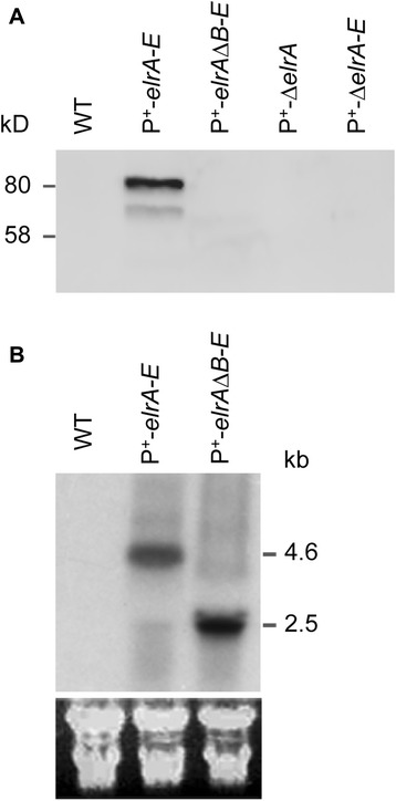
Fig. 2.

Detection of ElrA protein and elr transcript. a) Western blot analysis of total protein extracts from WT and mutant strains of E. faecalis, that was performed using a 12 % SDS-PAGE and polyclonal rat anti-ElrA antibodies, is shown. Band at ~80kD corresponds to the predicted size of ElrA, whereas the additional band represents a degradation product. b) Northern blot analysis of elr operon performed with ~40 μg of total RNA which was extracted from exponentially growing cells. Names of strains analyzed are indicated at the top of each lane. Probes used were elrA-specific oligonucleotide probes. The estimated length of transcripts that agrees with their predicted sizes is shown on the right. Below, ribosomal RNAs were used as loading controls
