
Fig. 3.
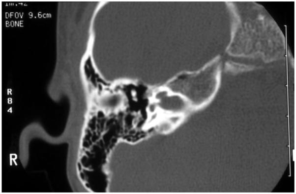
High-resolution computed tomography of the temporal bone, with axial cuts of the right ear at the level of the modiolus, of a boy with mixed hearing loss: the cochlear canal is enlarged creating abnormal patency between the cochlear fluids and the subarachnoid space. This child has X-linked mixed hearing loss DFN4.