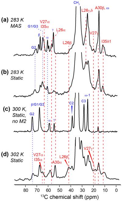
Figure 4.
1D 13C CP spectra of DMPC/DHPC bicelles with and without M2TM-AH under static and MAS conditions. (a) 13C CP spectrum of peptide-bound bicelles at 283 K under MAS, showing resolved protein (red) and lipid (blue) signals at isotropic 13C chemical shifts. (b) Static 13C spectrum of peptide-bound bicelles at 283 K. The bicelle is in the isotropic phase at this temperature, thus the chemical shifts are the same as in the MAS spectrum but the linewidths are broader. (c) 13C static spectrum of oriented bicelles without M2 at 300 K. The lipid chemical shifts differ from those in the MAS spectrum in (a) due to the presence of CSA under the oriented condition. (d) Static 13C spectrum of M2-bound oriented bicelles at 302 K. The peptide 13C chemical shifts are slightly different from the isotropic shifts in (a) and (b) due to the presence of CSA. Blue and red dashed lines guide the eye for chemical shift changes under different experimental conditions.

