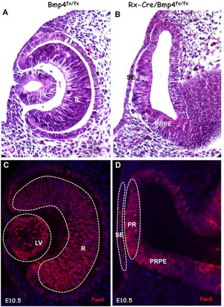
Figure 1.
Sections of the optic cup and lens in E10.5 wild type eyes and the optic vesicles and surface ectoderm from Bmp4CKO embryos. Embryos were littermates. A. An H&E-stained wild type eye with lens vesicle and optic cup. B. An H&E-stained Bmp4CKO embryo in which the lens did not form and the optic cup did not invaginate. C. A wild type eye stained for the transcription factor Pax6. Dotted lines around the lens and retina indicate the tissues that were laser microdissected for microarray analysis. D. A Bmp4CKO embryo stained for Pax6. The dotted lines indicate the tissues that were laser microdissected for microarray analysis. For all embryos, dorsal is toward the top of the figure. LV, lens vesicle; R, retina; RPE, retinal pigmented epithelium; SE, surface ectoderm; PR, prospective retina; PRPE, prospective retinal pigmented epithelium.

