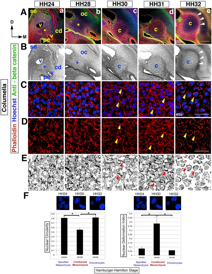
Fig 1. Dynamic cell shape changes reveal the timing of the columella.
(Rows A,B) Overview of columella condensation in the context of surrounding tissues, from HH24-32 in transverse section at the 2nd pharyngeal arch level, left side of the head (20x). Dorsal (D) is to the top, the midline (M) is to the right and surface ectoderm (se) is to the left. (A) Hoechst (blue)—nuclear DNA, Phalloidin (red)—F-actin, anti-beta catenin (green)—cell membranes and epithelia. (B) Black and white view. (Rows A,B) An asterisk indicates the position of nascent columella and the overlying nascent otic capsule (oc), from HH24-28. The nascent columella is surrounded by the cochlear duct (cd), pharyngeal endoderm (pe) and blood vessels (v). At HH30, the columella (c) condenses and by HH32 there is overt chondrocyte differentiation in the columella and overlying crescent shaped otic capsule. The perichondrium (arrowheads) surrounding the columella is apparent. (Rows C-E) F-actin rearrangements during columella condensation are apparent at higher magnification (60x). F-actin in red and nuclei in blue, with black and white images highlighting F-actin rearrangements and nuclei (row E). (Rows C-E) HH24-28, F-actin is disorganized and nuclei rounded in shape. At HH30/31, cell-cell actin bridges form (arrowheads), cells adopt rhomboid shapes and concomitantly distort nuclear shape. At HH32, overt differentiation is observed with cell-cell actin bridges replaced by cell-ECM adhesions leading to stellate shaped cells with rounded nuclei. (F) Quantification of nuclear circularity and deformation index during condensation and overt chondrogenesis. At HH24, nuclei are rounded with little distortion; in contrast, at HH30 during condensation, there is significant loss of nuclear circularity and a significant increase in the deformation index. This reverses during overt differentiation of chondrocytes when the nuclei of the now stellate shaped cells become rounded once more. Asterisk indicates p value <0.05. Abbreviations: cd-cochlear duct, c-columella, D-dorsal, M-midline, oc- otic capsule, pe-pharyngeal endoderm, se-surface ectoderm and v-blood vessel. Scale bars represent 150 μm (20x) and 25 μm (60x).

