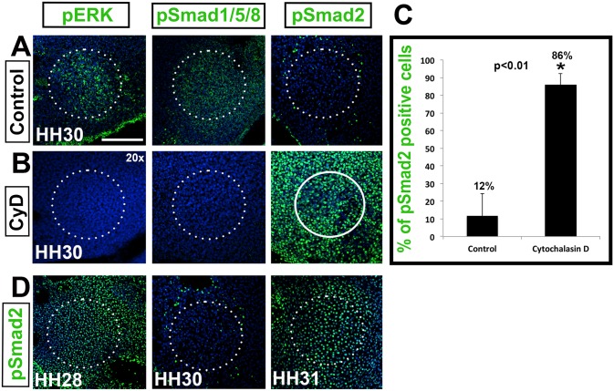
Fig 5. Actin reorganization is required for columella condensation.
(A,B) Tissue slices treated with Cytochalasin D. The activation status of FGF, BMP and TGF-β signaling pathways in the columella (circled) as measured by pERK, pSmad1/5/8 and pSmad2 (green) immunolabeling, respectively, at HH30. (B) Cytochalasin D treatment reverses the activated signaling patterns. (C) Graph of pSmad2 positive cells at HH30. Compared to 12% of control cells, 86% of cells remained pSmad2 positive in treated explants (asterisk, p<0.01). (D) TGF-β signaling in control tissue, showing the normal expression of TGF-β between HH28 and HH31. pSmad2 immunolabeling shows that TGF-β signaling is high at HH28 before condensation, down regulated at HH30 during condensation, and up regulated at HH31 following condensation. Scale bar represents 75 μm (20x).

