Fig. 5.
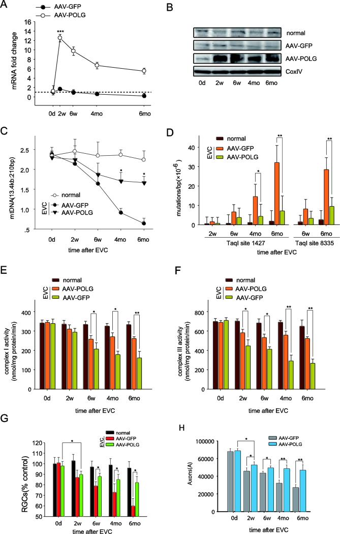
Intravitreal injection of AAV2-POLG prevented mtDNA alterations and promoted RGC survival in the rat glaucomatous model. A, POLG mRNA levels in the isolated RGCs were quantified by real-time PCR at 0 days, 2 weeks, 6 weeks, 4 months, and 6 months after EVC with AAV2-POLG/GFP. The fold change relative to normal age-matched controls was calculated using the ΔΔCt method. B, The mitochondrial protein levels of POLG in the RGCs were detected by western blot at different time points after EVC with AAV2-POLG/GFP. CoxIV was used as an internal reference. C, The mitochondrial DNA damage in the isolated RGCs was determined by LX-PCR after EVC with AAV2-POLG/GFP. D, The random mutation capture assay revealed that the point mutation frequency in mtDNA decreased at either position 1,427 or 8,335 of the mitochondrial genome after EVC with AAV2-POLG/GFP. E, F, The measurement of complex I (E) and III (F) activities in mitochondria isolated from RGCs at different times after EVC with AAV2-POLG/GFP. G, H, Quantitation of DiI-labeled RGCs (G) and toluidine blue-stained axons (H) showed that RGCs and their axon survival increased in EVC-treated eyes with AAV2-POLG compared to those with AAV2-GFP. n=24 retinas/time point/group (A, B, C, D). n=21–23/ time point/group (E, F). n=6/ time point/group (G, H).
For each, the values are the means ± SEMs. *p<0.05, ** p<0.01 and ***p<0.001 compared with the EVC-treated eyes with AAV2-GFP. normal: age-matched normal control. EVC: episcleral vein cauterization. w: week, mo: month.

