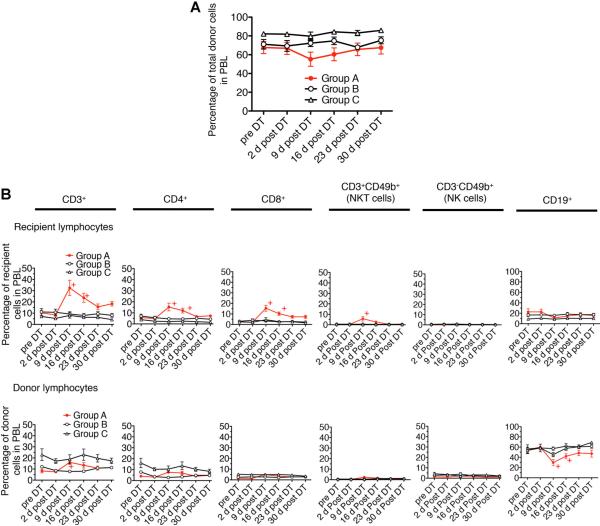
Figure 4. Peripheral blood lymphocyte (PBL) levels of donor chimerism were preserved after Foxp3+ cell depletion.
(A) PBL were stained with anti H-2Dd (donor MHC class I) mAb and plotted over 30 days after diphtheria toxin (DT) treatment. The difference between three groups is statistically insignificant by analysis of variance (p = 0.116). (B) Analysis of lymphocyte subpopulations after DT treatment shows a relative increase in recipient-derived T cells (CD4+, CD8+ and NKT cells) and a corresponding relative decrease in donor-derived B cells (CD19+ cells). PBL were stained with anti-CD3, CD4, CD8, CD49b (Dx5) and CD19 mAb. The graph in A shows the percentage of donor cells in PBL following DT treatment. The graph in B shows the percentage of recipient or donor-derived subpopulations in the whole peripheral blood lymphocytes. +p < 0.05 compared with data in Group A at pre-DT (t-test). Results represent mean values ± s.e.m. of 12, 6 and 5 recipients in Groups A, B and C, respectively.

